


Neuron
What is a Neuron?
The neuron is a nerve cell that is a specialized cell in the transmitting of nerve impulses. Neurons used to send signals to the brain, muscles, and glands. There are about 100 billion neurons in the brain to send signals. The signals transfer by neurons help the different parts of the body to communicate with each other. The neurons send chemical signals known as ‘neurotransmitters’ which works quickly for the reaction.
Neuron Diagram:
Neuron structure is described below :

What does a Neuron look like?
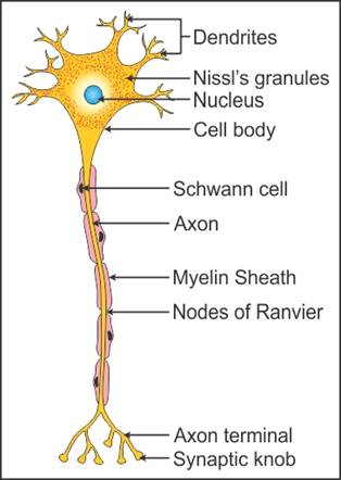
The neurons consist of a large number of extensions, which are known as Dendrites. The meaning of dendrite is ‘branches of the tree’. They usually look like the branches of trees or spikes extending out from the body of the cell. It can also say that the neuron looks like a ‘spider web’. The primary surfaces of dendrites receive chemical signals or neurotransmitters from the neuron and transfer to the other neuron.
Neuron structure
Axons:
The axons are the thread-like parts of nerve cells across which the impulses conduct from one cell body to the other. Axon is like a cable along with each neuron away from the main part of the cell body. It is many times thinner than a human hair and used to travel electric impulses from the neuron to away to receive by the other one.
Dendrite:
The dendrite is the extension of short branches of the neuron or nerve cell. Along the dendrites, the impulses received from other cells at the synapses which are transmitted to the cell body. The dendrite has a branching tree-like structure. It used to send and receive signals from one neuron to the other.
Spine:
The spine is a series of vertebrae that are extending from the skull to the back by enclosing the spinal cord. It provides support to the thorax and abdomen. The spine is the column of bone which used to surround and protect the spinal cord. The spine can be categorized by the body level i.e. cervical spine (neck), thoracic spine (upper and middle back), and lumbar spine (lower back).
Action Potential:
The action potential is the change of electric potential which is associated with the passage of an impulse across the muscle cell membrane or nerve cell membrane. The action potential works as a temporary shift, such as from negative to positive charge), in the membrane of neurons, the potential caused by ions flow in or out of the neuron.
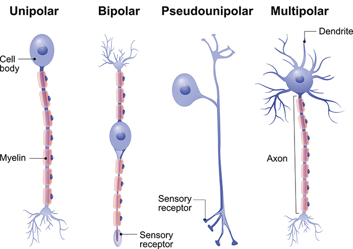
Types of Neurons
There are many varieties of neurons in your body. Each kind is specialized to be sensible at doing various things.
1. Multipolar nerve cell
Multipolar neurons have one axone and lots of dendritic branches. These carry signals from the central systema nervosum to alternative components of your body like your muscles and glands.
2. Unipolar nerve cell
Unipolar neurons are called sensory neurons. They need one axone and one dendrite branching off in opposite directions from the cell body. These cells pass signals from the outer surface of your body, like touch, along with the central systema nervosum.
3. Bipolar nerve cell
Bipolar neurons have one axon and just one dendrite branch. They pass signals from one nerve cell to the next subsequent within the central systema nervosum.
4. Pyramidal nerve cell
Pyramidal neurons are named on the basis of the form of their cell body, which appears like a pyramid. They need one axone and two main dendrite branches. These cells pass signals within the brain and tell your muscles to move.
5. Purkinje nerve cell
Purkinje neurons (named after the person who discovered them) are found within the cerebellum, the part of the brain that controls balance, coordination, and temporal order of actions. They need one axone and a really dense and complex dendrite arrangement.
Sensory Neuron
Sensory neurons are nerve cells inside the systema nervosum accountable for converting external stimuli from the organism’s surroundings into internal electrical impulses.
For example, some sensory neurons reply to tactile stimuli and may activate motor neurons in order to attain muscular contraction.
Such connections between sensory and motor neurons underlie motor reflex loops and several other varieties of involuntary behavior, together with pain turning away.
In humans, such reflex circuits are normally placed within the medulla spinalis.
Motor neurons
Motor neurons of the medulla spinalis are a part of the central nervous system (CNS) and connect with muscles, glands, and organs throughout the body. These neurons transmit impulses from the neural structure to skeletal and smooth muscles (such as those in your stomach) and help indirect management of all of our muscle movements.
There are in reality two kinds of motor neurons: those who travel from spinal cord to muscle are referred to as lower motor neurons, whereas those who travel between the brain and neural structure are known as higher motor neurons.
Interneurons
As the name suggests, interneurons are those in-between – they connect the spinal motor and sensory neurons. Additionally, as transferring signals between sensory and motor neurons, interneurons may also communicate with one another, forming circuits of assorted complexness. They’re multipolar, similar to motor neurons.
