


Skeletal System
Overview:
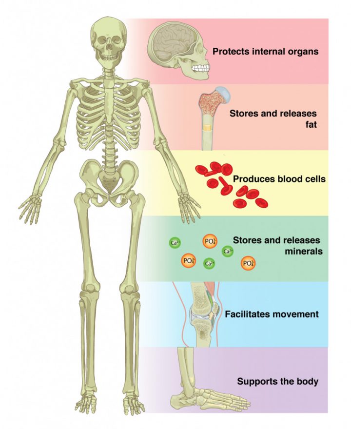
Skeletal System defines as an organ system which gives the ability to body for movement, shape, and support. The skeletal system makes a structure or framework of all bones, or rough rigid material which support the body of animals, or humans. The musculoskeletal system is made up of bones, cartilage, muscles, ligaments, tendons, joints, and other connective tissues which perform several functions such as support body, movement, and protects the internal organs from external damage. The skeletal system also performs the function of the main storage system for minerals.
The skeletal system is also responsible for the production of blood cells which occurs in red marrow and yellow marrow. The Skeletal system serves as the main storage system for some minerals, calcium, and phosphate. These minerals stored in bones, when the amount of minerals is high. While the amount of minerals is low, it withdraws from bones.
A newborn baby has 305 bones in his skeleton system which is made by cartilage. when he grew up, his skeletal system structured properly, and many bones attach with each other and then became a skeletal structure of 206 bones.
Skeletal System Anatomy
The skeletal system of a mature individual is composed of 206 individuals. These skeletal system bones are organized into two major divisions:
- Axial skeleton
- Appendicular skeleton.

Basically, the Axial skeleton of the skeletal system runs along the body’s middle axis and it is comprised of totally 80 bones in the following regions:
- Skull
- Hyoid
- Auditory ossicles
- Ribs
- Sternum
- Vertebral column
While the Appendicular skeleton is composed of 126 bones in the subsequent regions:
- Upper limbs
- Lower limbs
- Pelvic girdle
- Pectoral (shoulder) girdle
Skull
The skull is basically composed of 22 bones which are merged together except for the bones of Mandible. This twenty-one (21) united bones are separate in kids to allow the skull and brain to grow, while later on fuse to provide additional strength and protection as an adult. The mandibular bone remains as a movable jaw bone and forms the only movable joint within the skull with the temporal bone.
The bones of the superior portion of the skull are called the cranium and shield the brain from injury. The bones of the inferior and anterior portion of the skull are called facial bones and support the eyes, nose, and mouth.
Hyoid and auditory Ossicles
The hyoid maybe a little, U-shaped bone found in inferior to the mandible. The hyoid is that the only one bone within the body that doesn’t create a joint with the other bone—it may be a floating bone. The hyoid’s main function is to assist hold the trachea open and to create a bony connection or relation for the tongue muscles.
The malleus, incus, and stapes—known are jointly known as Auditory Ossicles. Basically, they are the tiniest bones within the body. Found in an exceedingly tiny cavity within the temporal bone, they serve to transmit and amplify sound from the eardrum to the inner ear.
Vertebrae
Within the human body, there are Twenty-six vertebrae create the Vertebral Column. They are termed by regions given below:
- Cervical (neck) – 7 vertebrae
- Thoracic (chest) – 12 vertebrae
- Lumbar (lower back) – 5 vertebrae
- Sacrum – 1 vertebra
- Coccyx (tailbone) – 1 vertebra
With the exclusion of the Sacrum and Coccyx, each vertebra is called for the initial letter of its region and its location along the Superior-Inferior Axis. For example, the first superior Thoracic Vertebra is known as T1 and while the most inferior Thoracic Vertebra is called T12.
Ribs and Sternum
The breastbone, or sternum, is a thin, knife-shaped bone that is situated on the midline of the anterior portion of the thoracic cavity of the human skeleton. The breastbone connects to the ribs by the help of thin bands of cartilage that are known as the Costal Cartilage.
There are twelve pairs of ribs that are present along with the breastbone which create the ribcage of the Thoracic region. The first seven ribs are called “True Ribs” and they connect the Thoracic vertebrae to the Breastbone through Costal cartilage. Ribs 8, 9, and 10 all are connected with the breastbone through cartilage that’s connected to the cartilage of the seventh rib, therefore we consider these to be “False Ribs.” Rib eleven and twelve are false ribs, and they are also considered to be “Floating ribs” because they do not have any gristle attachment to the breastbone from any direction.
Pectoral Girdle and Upper Limb
The pectoral girdle connects the higher limb (arm) bones to the axial skeleton and consists of the left and right clavicles and left and right scapulae.
The arm bone (Humerus) is that the bone arm region. It forms the ball and socket joint of the shoulder with the shoulder blade (scapula) and forms the elbow joint with the lower arm bones. The radius and elbow bone (ulna) are the 2 bones of the forearm. The elbow bone (Ulna) is present on the medial part of the forearm and forms a hinge joint with the arm bone (humerus) at the elbow. The radius permits the forearm and hand to move over at the wrist joint.
The lower arm bones make the wrist joint with the carpals, a bunch of eight little bones that provide additional flexibility to the wrist joint. The carpals are connected to the 5 metacarpals that make the bones of the hand and that are connected with every one of the fingers. Every finger has 3 bones called phalanges, apart from the thumb, that only has 2 phalanges.
Pelvic Girdle and Lower Limb
Formed by the left and right hip bones, the pelvic arch connects the lower limb (leg) bones to the axial skeleton.
The leg bone (Femur) is the largest bone within the body and also the only one of the thigh (femoral) region. The leg bone (Femur) forms the ball and socket hip joint with the hip bone and forms the knee hinge joint with the shinbone (Tibia) and patella. Usually it is known as the kneecap, the patella is special bone because it’s only one of the bones that are not present at birth. The patella forms in early childhood to support the knee for walking and creeping.
The shinbone (Tibia) and leg bone (Fibula) are the bones of the lower leg. The shinbone (Tibia) is very larger than the leg bone (Fibula) and bears most of the body’s weight. The leg bone (Fibula) is principally a muscle attachment site and it is utilized to assist to maintain balance. The shinbone and leg bone (Fibula) make the ankle with the talus, one of the seven tarsal bones within the foot.
The tarsals are a bunch of seven little bones that create the posterior endpoint of the foot and heel. The tarsals make joints with the 5 long metatarsals of the foot. Then every one of the metatarsals forms a joint with one of the sets of phalanges within the toes. Every toe has 3 phalanges, apart from the large toe, that only has 2 phalanges.
Skeletal System Parts
Skeletal System is made up of different parts including bones, muscles, joints, and rough rigid parts. The skeletal system has two types of connective tissues; Tendons and Ligaments. Tendons works for the connection of bones to muscles and ligaments create a connection between bones and bones.
There are three main sections of the skeletal system:
- Bones
- Joints
- Cellular Composition
Bones
Our skeletal system has 206 bones, with more than 200 joints. These joints connect the bones with each other and cause many functions like bending, moving, etc. The skeletal system classified into two categories based on location:
- Appendicular Skeleton:
These bones are present in appendages like legs, arms, fingers, and toes.
- Axial Skeleton:
These bones lie towards the midline of the body that includes the skull, the rib cage, and the vertebral column.
Bones Division
Types of Bones: Bones are divided into five parts based on shapes; long bones, short bones, flat bones, irregular bones, and sesamoid bones.
- Long Bones:
These bones are longer in length than width. These bones are shaft and have two rounded ends. These bones are generally lying in the limbs, femur.
- Short Bones:
These bones are short in length having the same width and length. These bones have a cube-like shape. These bones lie in carpals and tarsals.
- Flat Bones:
These bones are thin and flat and may not have all of them completely flat shape. They protect internal organs by attaching with the surface area of muscles. These bones are scapula and sternum.
- Irregular Bones:
These bones have an irregular shape and complex shape and may have rigid, long, and short surfaces. Examples of these bones are vertebrae and many skull bones.
- Sesamoid Bones:
These bones have a similar shape to sesamoid seed. Structure of these bones is flat and small. These bones lie in knee and joints.
Joints
The bones are connected with each other with the help of joints. The skeletal system is made by different types of joints; fibrous, cartilaginous, and synovial. Every joint has a particular method of functions.
- Fibrous Joint:
The fibrous joints are fixed and immovable in the skeletal system as sutures of the skull. The coronal suture makes the connection between parietal and frontal skull bones. The sagittal suture creates a connection between two parietal bones. The lambdoid suture connects the parietal with occipital.
- Cartilaginous Joint:
These joints are moveable joints, which contains symphyses or synchondroses joints. These bones are the rib cage and spinal column.
- Synovial Joint:
The synovial joints connect two bones, which contains cartilage lined cavity filled by fluids. These joints are also known as Diarthrosis joints, which are more flexible joints between bones. These bones have not physically connected with each other. It can move freely in relation to each other.
Cellular Composition
The cellular composition in the form of cells which makes the bone matrix. The cell which makes bone matrix called Osteoblasts. And the mature bone cells are known as Osteocytes. Osteoclasts are the special kind of cells which removes bone matrix that lies during bone remodeling. These tissues are gigantic cells.
Microscopic Structure of Bones
The skeleton makes up approximately 30-40% of an adult’s body mass. The skeleton’s mass is formed by the dead bone matrix and lots of small bone cells. Roughly 1/2 the bone matrix’s mass is water, whereas the other half is albuminoid protein and solid crystals of CaC03 and inorganic phosphate in the form of calcium phosphate.
Living bone cells are found on the sides of bones and in little cavities inside the bone matrix. while these cells generate little of the overall bone mass, they comprised of many important roles within the functions of the skeleton. The bone cells enable bones to:
- Grow and mature.
- Be mended the bones at the point of injury.
- Being able to keep minerals inside the bones
Skeletal System Structure
Bone tissue in the skeletal system is made up of two kinds of tissues:
- Compact bone
- Spongy bone.
Compact bone is naturally compact and hard and always lies outside of the bone. The connective tissue which lies on the outside of the bone called Periosteum. The periosteum is made by cellular and fibrous tissues which help in the attachment of muscles and joints. The connective tissues which line the marrow cavity are known as Endosteum. The diaphysis is the shaft of the bone and its swollen ends are known as the epiphysis. The bone matrix tissue is made by two components, which are organic and inorganic part. The organic parts consist of fibers and inorganic part contains the minerals.
Skeletal System Diseases
There are some diseases which affect the skeletal system severely. Some of these diseases are described as below:
The curvature of the Spine:
The curvature of the Spine occurs when the spine cannot curve its usual shape. Commonly, the spine follows toward and backward curves.
The curvature of the Spine has three main kinds.
- Kyphosis:
It generates rounding curve in the upper back.
- Lordosis:
It generates lower back curve inward.
- Scoliosis:
It generates an S-shape or C-shaped curve in the spine.
Osteoporosis
Osteoporosis is a disease of the skeletal system which causes weakness of bones. In this, the body loses too much bone, which makes bones little. Bones may break from fall, or in some serious cases, from minor bumps or sneezing. Osteoporosis means the Porous bones, in which healthy bone looks like a honeycomb.
Arthritis
Arthritis is joint inflammation. Pain and a limited range of movement are caused by Arthritis. Many things may cause this disease as cartilage breakdown, autoimmune conditions, or infection.
Skeletal System Functions:
Interesting Facts about the Skeletal System
- The skeletal system has 206 different kinds of bones.
- The human skeleton made by two kinds of bones; Cortical and Trabecular.
- Bone marrow is the spongy material inside of large bones.
- Babies have around 305 bones by birth.
- As the baby grew up, 305 bones reduce to 206 bones.
- The inner air has the smallest bone known as Staples.
- The thigh bone is the largest and longest bone of the skeleton.
- Three bones are connected at knee joint; femur, tibia, and patella.
- Osteoporosis is characterized by brittle bones.
- Each hand is made by 14 phalanges, 5 metatarsals, 5 tarsals, calcaneus, and talus.
- The skull is made by 22 different bones which grow in size together.
- Bones are consists of the mineral mixture, organic matrix, water, and lipids.