


Gram Staining
Introduction of Gram Staining
What is Gram Staining: Dr. Christian Gram in 1884 introduces Gram staining technique and classified bacteria on its Gram characters which might be Gram-negative or Gram-positive. In the microbiology field, extensive staining techniques are used. In accumulation, these staining techniques also help in the determination of cell size, structure, and configuration. It’s a sort of identification test.
Gram Staining Principle
Here are the following main principles of gram staining techniques.
- Cell wall structure identifies either cell is Gram-positive or negative in nature.
- During the procedure when we stained by primary stain and secure it by a mordant.
- At that time, some bacteria have the ability to hold or retain the primary stain by counterattacking decolorization.
- While some bacteria decolorized by the use of a decolorizer. The bacteria which retain its primary stain are called Gram-positive and the bacteria which will be decolorized called Gram-Negative.
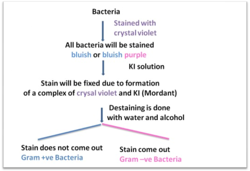
- Crystal violet (CV), when dissolved in water, make CV+ and Cl– ions in its solutions. These ions infiltrate by a cell wall and cell membrane. This happens in both Gram-negative and Gram-positive cells. The CV+ ion interrelates with (-) negative charge bacterial cells components and gives a purple stain to the cell.
- Iodine (I), is practice as a mordant that intermingles with CV+ cells and produces large multiplexes of violet crystal.
- When alcohol or acetone as decolorizer is added it reacts with cell membrane lipids.
- Gram-negative cells have very thin 1-2 layers of peptidoglycan and have a lipopolysaccharide layer which is dissolved by adding Alcohol.
- Due to this ability gram-negative cells unable to recollect the complexes and were decolorize when the complex is washed away completely.
- In divergence, Gram-positive celled organisms by Ethanol treatment renovates desiccated. Due to this cell wall pores closed. and due to this stain cannot exist in the cell.
- After this decolorization procedure, Gram-positive celled retained its purple and in contrast, the Gram-negative celled lost its purple color.
Gram Staining Reagents
Gram Staining Kit
-
Primary Stain: Crystal Violet
Solution A :
Ethyl Alcohol= 20 ml
Crystal Violet = 2 gm
Solution B :
Distilled Water = 80 ml
Ammonium Oxalate = 0.8 gm
After this mix A and B solution. Keep this solution for 24 hours and then filter it. Place in the yellowish-brown painted bottle.
-
Mordant: Gram’s Iodine Solution
Potassium Iodide = 2 gm
Iodine = 1 gm
Distilled water = to 100 ml
Mix them all and stockpile in a yellowish-brown colored bottle.
-
Decolorizer: 95% Ethanol or 1:1 Acetone with Ethanol
Ethanol (95%) = 50ml
Acetone = 50 ml
-
Counterstain: Safranin
Safranin O = 0.34 gm
Distilled water = 90ml
Absolute alcohol = 10ml
Mix them all and filter it and store it in a yellowish-brown colored bottle.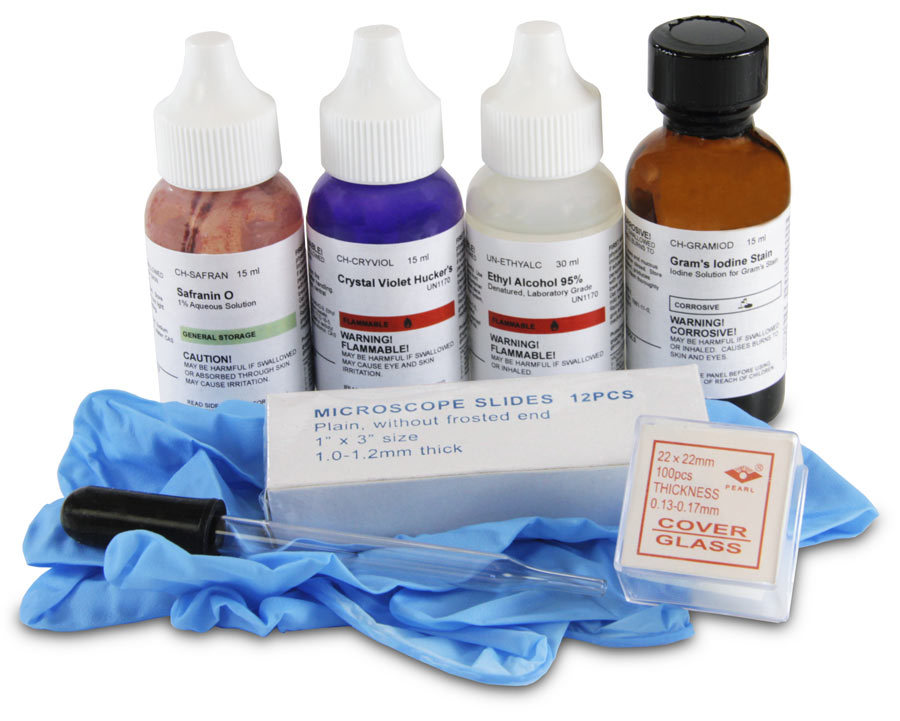
Gram Staining Protocol
Smear preparation:
- First of all, take a lubricant permitted dry slide.
- Take the inoculating loop and disinfect this inoculating loop by the heating it on the Bunsen burner flame.
- Later on the transference of a loopful culture with the help of antiseptic loop and make a slur at the epicenter.
- Smear neither reedy or nor very profuse.
- Consent the smear to parched in the Air. Fix the gasping smear on the slide by transient the slide 3-4 times through the flame quickly with the smear side facing up.
 You May Also Like: Virus Structure | Definition | Classification & Characteristics
Gram Staining
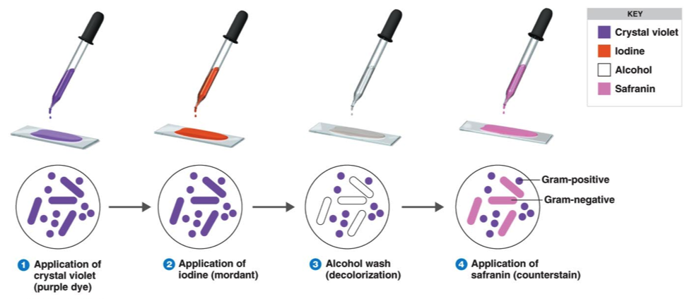
- Dwelling the slides on the special rods which are used for staining.
- Refuge the smear with the stain of crystal violet and consent for 1 minute.
- Rinse prudently below tap water.
- Inundation the smear with solution Iodine and consent it for one minute.
- Trench off the iodine gram solution.
- Rinse the slide again in tap water.
- Deluge the slide by a decolorizing agent.
- Wait for 20-30 seconds.
- This process is also performed by accumulating drop by drop on the slide.
- Continue this process until the decolorizing agent seriatim from slides.
- Moderately wash the slide by water and trench it wholly.
- Counterstain is added with Safranin.
- After this wait for almost 30 seconds to 1 minute.
- Rinse the slide in the ancillary tap water stream.
- Continue this process till no color seems in the seepage
- Dry the slide by blotting paper.
- After this observe the slide beneath the Microscope.

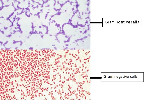
Gram Staining Results
The staining outcomes are as follows :
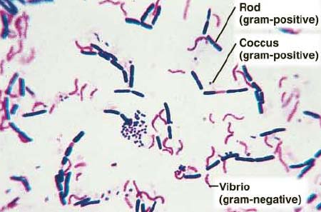
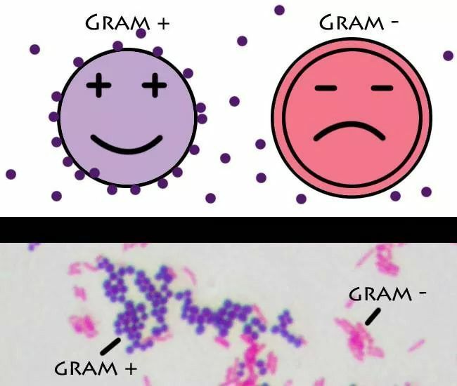
Gram-Positive Cell: Dark Purple color seemed
Gram-Negative: Pale to dark Red color seemed
Yeasts cell: Dark purple color
Epithelial cells: Pale red color
You May Also Like: Functions of Lipids | Definition | Classification | Examples
Gram Stain Result Interpretation
Gram Positive and Gram Negative Bacteria List
|
Gram-Positive Bacteria |
Bacillus Species | Clostridium Species | Corynebacterium Species
|
| Bacillus anthracis
Bacillus cereus
Bacillus subtilis
|
Clostridium difficile
Clostridium perfringens
Clostridium tetani
|
Corynebacterium diphtheria
Corynebacterium jeikeium
Corynebacterium urealyticum |
|
Gram-Negative Bacteria |
Brucella Species |
Haemophilus Species:
|
Neisseria species:
|
| Brucella abortus
Brucella canis Brucella melitensis Brucella suis |
Haemophilus influenzae
Haemophilus ducreyi Haemophilus avium |
Neisseria gonorrhoeae Neisseria meningitidis |