Renal Tubule
Renal Tubules are the major part of the Nephron. When blood leaves the Renal Corpuscle after this filtrate passed by the special tubules called Renal Tubule.

Kidney Tubules
Kidney Tubules consists of the following parts :
- Proximal Convoluted Tubule
- Loop of Henle.
- Distal Convoluted Tubule.
- Collecting tubule.
- Collecting duct.
Proximal Convoluted Tubule
These are the initial helical section of the Renal Tubule that gets the filtrate by the Glomerulus and Renal Corpuscle. Proximal Convoluted Tubules crinkled by an Epithelium Simple Cuboidal cells. These cells make exploit the number of transporter proteins in the inside layer of cell membranes. The total length of the Proximal Convoluted Tubule is ~14 mm. It is the broadest portion of the Renal Tubule and due to this, it provides the most important location for tubular filtrate reabsorption. Approximately ~80% of the filtrate water, nutrients, electrolytes, small proteins, albumin molecules are reabsorbed. These tubules play a minor role in the tubular secretion of Nitrogenous wastes and Electrolytes. The functioning of the Proximal Convoluted Tubules is regulated by Autoregulatory Mechanisms. This mechanism is reactive to a variety of hormones including Aldosterone, Atrial Natriuretic Peptide, and Parathyroid hormone.
Loop of Henle
It is basically Nephron Loop also called Loop of the Nephron. It is the second part of Renal Tubule which is normally of U-shaped. It is situated sandwiched between the Proximal and Distal Convoluted Tubules. Inside the Kidney, it is located in the Kidney’s Cortex or might be encompassed into the Renal Pyramid and inside the Medulla. Loop of Henle is the thinnest portion of the Renal tubule comprising of slighter lumen inside them. This segment is lined by a single cell layer of squamous epithelium cells. These cells play a vital role in the ions and water transport and help in concentrating the urine. It is the chief locations of facultative water reabsorption. This portion of the Renal Tubule is highly reactive or responsive to Antidiuretic Hormone = ADH which is also called Vasopressin.
Nephron Loop
Nephron Loop consists of two parts whose brief description is described below:
- Descending Loop of Nephron
- Ascending Loop of Nephron
Descending Limb of the Loop of the Nephron
The Proximal Portion of the Loop of the Nephron help in the transportation of the filtrate from the Kidney’s Cortex down into the Medulla portion. This limb is lined by squamous cells which are extra permeable to water and slightly less permeable to Chloride and Sodium. Due to this, water tends to leave the descending limb by the process of Osmosis and enter into the Medulla interstitial fluid. From this, it enters into the Venous Capillary Drainage and then back into the Systemic Circulation.
Ascending Limb of the Loop of the Nephron
The Ascending limb which helps in the transportations of the filtrate from the Kidney’s medulla back to the Renal Cortex. This limb is lined by squamous cells and these cells are less permeable to water and extra permeable to Chloride and sodium ions. Due to this Sodium-Potassium ATPase pumps, sodium ions are taken out from the filtrate and these ions are replaced by Potassium ions in the filtrate.
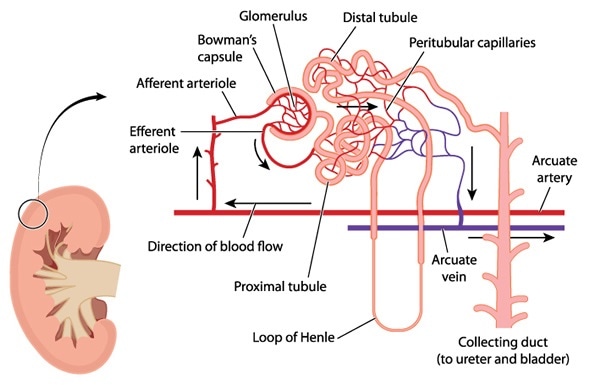
Distal Convoluted Tubule
It is the last coiled portion of Renal Tubule which accepts the filtrate by the loop of the Henle. These tubules are wizened by cuboidal epithelium cells. While they lack a brush border as Proximal Convoluted Tubules have. These tubules allow less reabsorption almost about <20% of water, nutrients, electrolytes, small proteins, e.g., albumin, are reabsorbed. They play a vital role in the secretion of various electrolytes, particularly H+ ions, and nitrogenous wastes compounds. They also help in the removal of “toxins,” by plasma and actively transport them into the Urine. So that they are expelled out from the body. All this mechanism is controlled by Autoregulatory Mechanisms. That mechanism is very responsive to various hormones for example Antidiuretic hormone = ADH = vasopressin, Angiotensin II, Aldosterone, and Atrial Natriuretic Peptide. These tubules at the end empty urine into the Collecting ducts.
Collecting Duct
It is the basically last portion of renal tubules in which urine is collected from the Distal Convoluted Tubules.
Collecting Duct Function
- Collecting Duct transport that urine to the Papillary Ducts. That is present at the tips of Medulla of the Renal Pyramids.
- From where the urine conceded into the minor calyxes.
- This area is lined by cuboidal to simple columnar epithelial cells which help sodium chloride and urea actively transported from the urine into the Medullary Interstitial Space that preserves the osmotic gradient among the Renal Cortex and the Renal Medulla.
Renal Corpuscle Diagram

Renal Corpuscle
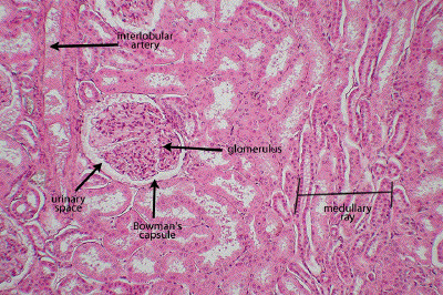
The renal corpuscle is also known as Malpighian body of the Nephron. It is the filtration element of vertebrate nephrons and efficient units of the kidney. Renal Corpuscle comprised of capillaries knot called glomerulus which is bounded by a double-walled capsule which is known as Bowman’s capsule that unlocks into a tubule. Blood pressure services in transfer of macromolecules (e.g., proteins) by glomerular capillaries into the Bowman’s capsule. This remainder is called capsular urine, at that time passes into the tubule for further processing.
Renal Corpuscle Histology