What is Microscopy?(Introduction to microscopes and how they work)
Introduction to Microscopy: The cells are very minuscule and multifaceted living groups. The cell has small magnitudes and translucent nature and appearance which create problems for cell biologists that are trying to comprehend its construction and functioning. For this purpose, various instruments and techniques have been developed to study the structure of the cell, cells molecular organization and function.

Introduction to Microscopy
A (microscopy) microscope (from the Ancient Greek: μικρός, mikrós, “small” and σκοπεῖν, skopeîn, “to look” or “see”) is an instrument that amplifies objects until they are too small to be seen by the naked eye. Microscope creates an image in which the object appears bigger. Photographs of cells that are taken using a microscope and these pictures can also be called micrographs.

The microscopy must complete three tasks during functioning :
- Microscopy produces a magnified image of the specimen that is not seen by the naked eye.
- Microscopy discrete all the important details in the micrograph
- Microscopy purifies the details that are not visible to the human eye or camera. The microscope is specially designed to get a magnified image. It has multiple-lens designs with objectives and condensers. But in ancient time very simple single lens devices that are known as a magnifying glass.
Commonly the diameters of the majority of cells are ranged from (5-500 µm). But maximum cells diameter is between 10-150 µm. The system International (SI) units of length are
- 1 meter (m) = 1000 millimetres (mm)
- 1 mm (10-3m) = 1000 micrometres (µm)
- 1 µm (10-6m) = 1000 nanometres (nm)
- 1 nm (10-9m) = 1000 picometres (pm)
Olden times microscope (Ancient Microscopy):

Microscopes are instruments premeditated to produce magnified photographic images of small objects that are not seen through the naked eye. In 1960s British microscopist, Robert Hooke stunningly constructed a microscope. This microscope consists of an objective lens. the objective lens is located near the specimen and maintained focus by turning the body of the microscope to move the object closer to or beyond from the specimen. This microscope also consists of an eyepiece lens that is placed on the topmost side of the microscope. The microscope also has a field lens within the barrel according to the need.
You May Also Like: Introduction to Cell
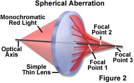
The microscope in the figure that is formulated by Robert Hooke is illuminated through the oil lamp and water-filled spherical reservoir. Light from the spot lamp is diffused when it permits through the reservoir and it is then fixated on-to the specimen with a lens that is directly attached to the reservoir. But there is a problem while using this microscope. This microscope is tormented from chromatic (and spherical) aberration, and all images observed in white light contained “halos” that were either blue or red in color.