


The Human Eye
Eye Definition
In Human, Eye is a dedicated intellectual organ that is proficient of receipt Photographic images, which are then passed toward the Brain. The Human Eye is a body part which permits image. Rod and cone cells in the retina allow conscious light perception and vision including color differentiation and the perception of depth. Eye Anatomy is described below:
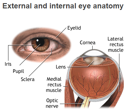
Human Eye Structure
Image Of Human Eye

Diagram of Human Eye with Labelling
Eye Anatomy
Complete Physiology of Eye is described below in the given paragraph:
- The eye is rather like a living Camera. Each eye is a liquid-filled ball 2.5 cm in diameter.
- At the front of the eye is a clear, round window called the cornea. Behind the cornea is a “lens.
- A camera focuses by moving the lens nearer or further away from the object. The eyeball cannot be made longer or shorter. Instead, the eye focuses by changing the shape of the lens.
- The lens is elastic and its thickness is controlled by the ciliary muscles inside – the eye. This process is called accommodation.
- Together, the cornea and the lens behind it focus light onto a layer of sensory cells at the back of the eye. This layer is called the retina, and light makes chemical changes in the cells of the retina.
- These chemical changes set off nerve impulses which travel along the optic nerve to the brain. The image made on the retina is upside-down.
- Your brain has to correct the image so that you see it properly. Just behind the lens is a sheet of muscle called the iris. It is the colored part of the eye. In the center of it is a round hole, called the Pupil.
- When you look at a bright light, the Iris Muscles work to make Pupil smaller.
- This cuts down the amount of light entering the eye and so Protects the sensitive cells of Retina.
- If you are in the darkroom the Pupil gets bigger to allow maximum light to pass.
- The Iris’ muscles work automatically by the Reflex Action.
Eye Biology: The eyelids cover the eyes and protect them from damage, while the eyelashes flick away any dust approaching the eye. The tear glands produce tears, and when you blink this liquid is spread over the surface of the eye. It washes the surface of the eye and stops it drying out. Tears drain away through the tear ducts. Eye Black and White are very common.
Eye Anatomy and Function
Eye Parts and Functions are described below in the given table. The eye comprised of the following parts which produce clear Vision:
| Eye Parts | Description and Functions |
|---|---|
| Cornea | The Cornea is basically Eyes Outer covering. It is generally dome-shaped layer and its main function is to protect your eye from foreign substances which may damage the Eyes inner portion.
Cornea comprised of several layers which create a tough layer which provides additional support and protection. The cornea also helps the eye to properly focus on light more accurately. |
| Sclera | The Sclera has generally stated the “Whites” of the Eye.
This is a flat, white layer which is present on the outside, but the inside it is brown and comprises furrows that help the muscles of the eye to ascribe appropriately. The sclera delivers protection to the inner workings portion of the eye. |
| Pupil | The pupil is a black dot that presents in the middle portion of the eye.
This is actually a hole which takes in light so that the eye can focus easily. |
| Iris | The iris is the zone of the eye which comprises the pigment which springs the eye its shade. This area environs the Pupil and practices the dilator Pupillae muscles to expand or contract the Pupil. This permits the eye to take in extra or less light condition on how brightness is it around you. If it is too much brightness around you, the iris will shrink the pupil so that the eye can focus more efficiently. |
| Conjunctiva Glands | These are mucous layers that keep the exterior part of the eye saturated.
If the eye desiccates out it can become scratchy and painful. It can also become more vulnerable may lead to Infection. If the Conjunctiva Glands become infected the patient will tune its eye into “Pink Eye.” |
| Lacrimal Glands | Lacrimal glands are present on the Eyes outer corner. They are specialized for producing tears which help moisten or humid the eye whenever it becomes dry and also helps to flush out substances or particles which irritate the eyes. When tears flush out harmful irritant particles, it becomes more east to focus on properly objects. |
| Lens | The lens is present behind the Pupil. It is a clear layer which helps to focus the light which the Pupil takes in.
Lenses are placed by Ciliary Muscles, that help lens to deform its shape according to the light amount and intensity which hits on it. |
| Retina | The light which is focused by the lens alters on conveyed onto the Retina. The retina is made up of Rods and Cones which are arranged in specialized layers.
They transmit light into compounds and electrical pulsations. The retina is situated in eyes back, and it is also associated with the Optic Nerves. Optic Nerve will conduct the imaginings that the eye perceives to the brain so they can be interpreted. The Retina back is known as the Macula, which helps to understand the specifics of the object. The Macula center is, known as the Fova which will increase the feature of images. |
| Ciliary Body | The ciliary body is generally ring-shaped tissue that controls the Eyes lens movement. In other, it also controls the Lens shape. |
| Choroid | The choroid present among the retina and the sclera.
Its main function is to supply blood to the eye. |
| Vitreous Humor | The vitreous humor is the ointment or gel present in the Eyes backside. The main purpose of the gel is to provide accurate shape to the Eye. The gel revenues nutrients from the Ciliary body, Aqueous humor and the Retinal vessels due to this eye can persist healthily. |
| Aqueous Humor | The Aqueous Humor is a water-logged substance that fills the eye. It is divided into two chambers. The frontal chamber is located in anterior of the iris, and the posterior chamber is directly behind it. These layers permit the eye to preserve its shape. |
Eye Anatomy is very complicated.
External Eye Anatomy
Parts of The Outer Eye
Outer Eye portion comprised of the following parts:
- Eyelid
- Iris
- Pupil
- Sclera
Eye Function
Functions of Eyes are given below:
- Our eyes detect light. The two eyes are positioned so that each one gives a slightly different view of your surroundings. This gives us a Binocular Vision.
- Each eye produces an image, or picture, of what you are looking at.
- Information about the image in each eye is then sent along a nerve to your brain.
- The brain compares the images from each eye and makes a three-dimensional ‘picture’ of them.
- Your brain can also work out the size of an object and its distance from you.
- We need two eyes because with only one eye it is difficult to judge distance and depth.
- Both eyes can be moved in hallows or sockets in the skull. The movement is brought
- By muscles. You can control these muscles so that your eyes can look up or down or to the left or right.
Eyes and Brain
Inside each eye approximately about Million Nerve Fibers carry pictorial messages from the retina of the eye to the brain. The brain is basically the control center of the whole body. The brain combines the images what it actually sees. The brain also plays a vital role in the focused image on the Eye’s retina which is actually Upside Down, so that the Brain cracks pictures right adjacent up. This setback of the images is just like a mirror does in a camera. Inside the Eye, Glaucoma helps to increase pressure in the eye which confines the stream of compulsions to the Brain, triggering optic nerve damage.
Eyes Problems
Common Vision Problems are described below:
Eyesight Problems
Some people can see distant objects clearly but cannot focus on very near objects. They are said to be long-sighted. Other people can see near objects clearly but cannot focus on distant objects. They are short-sighted.
Long Sightedness ( Hypermetropia)
Long sight is caused by the eyeball being too short so that images are in focus behind the retina. This is quite common in older people. As people get older, the lens loses some of its elasticity and the ciliary muscles weaken so that the lens cannot be pulled thin. Long-sighted people can be helped when a convex lens is placed in front of each eye. The lenses help to focus light on the retina.
Short Sightedness (Myopia)
Short sight occurs when the eyeball is too long and images are focused in front of the retina. Short-sighted People can be helped if they wear spectacles or contact lenses that are concave.
Treatment for Common Vision Problems
Contact Lenses
Contact lenses are small, thin transparent discs that fit snugly over Cornea of the eye. Modern ones are made of soft, jelly-like plastic. The purpose of contact lenses is the same as that of spectacles to correct defects of eyesight. Many people find wearing spectacles is uncomfortable or difficult, especially when playing sports or during other activities. Contact lenses are lightweight, virtually invisible, they give a wide field of view and they do not steam up in wet weather.
Laser Eye Surgery
A laser produces a very narrow beam of light that is both powerful and delicate. It can be controlled very accurately. Surgeons can use a laser beam to make the shape of the surface of the cornea change slightly. The cornea then focuses images accurately onto the eye. After laser eye surgery, It is no longer necessary for the person to wear either spectacles or contact lenses
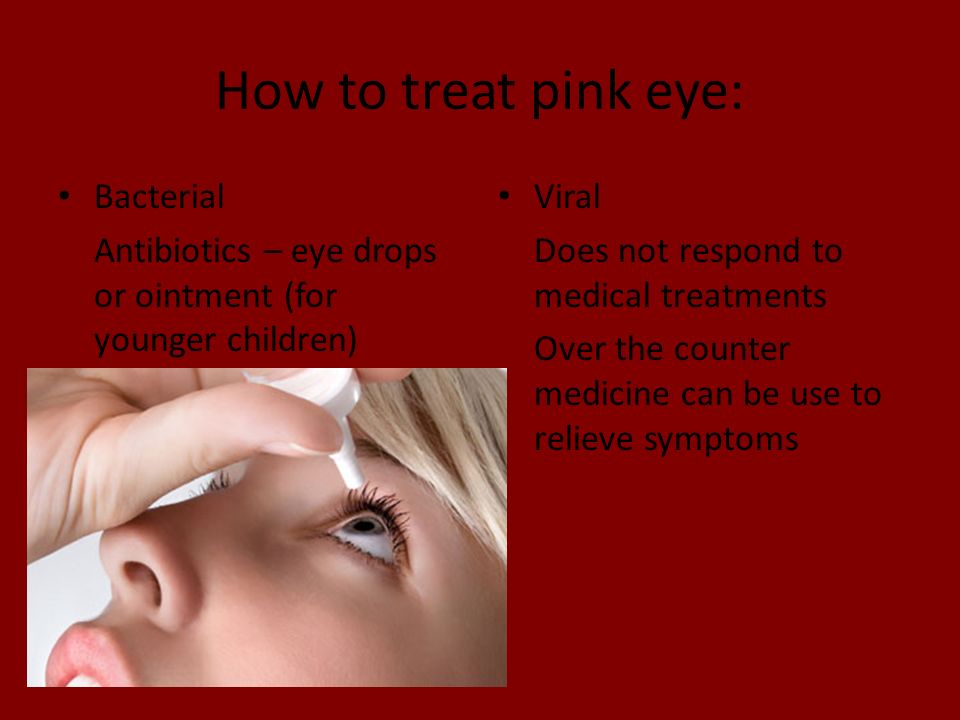
Pink Eyes

Pink Eye Treatment
Eye Anatomy is not easy to understand but o treat the Bacterial pink eye always use Antibiotic Eyedrops, ointment, or pills.
Eyelid Problems
Common Eyelid problems are given below:
- Eyelids which turn in or out
- Eyelids that wilt
- Abnormal blinking or jerking
Common Eye Infections
Common Eye Infections are described below:
- Conjunctivitis/Pink eye
- Keratitis
- Endophthalmitis
- Blepharitis
Rare Eye Diseases
Some Rare Eye Diseases are given below:
- Stargardt Disease
- Usher Syndrome
- Uveal Coloboma
- Retinitis Pigmentosa
- Retinoblastoma
- Anophthalmia and Microphthalmia
- Bietti’s Crystalline Dystrophy
- Behçet’s Disease
- Idiopathic Intracranial Hypertension
Eye Diseases That Cause Blindness
- Retinitis Pigmentosa
- Diabetic Retinopathy
- Cataract
- Glaucoma
- Age-Related Macular Degeneration
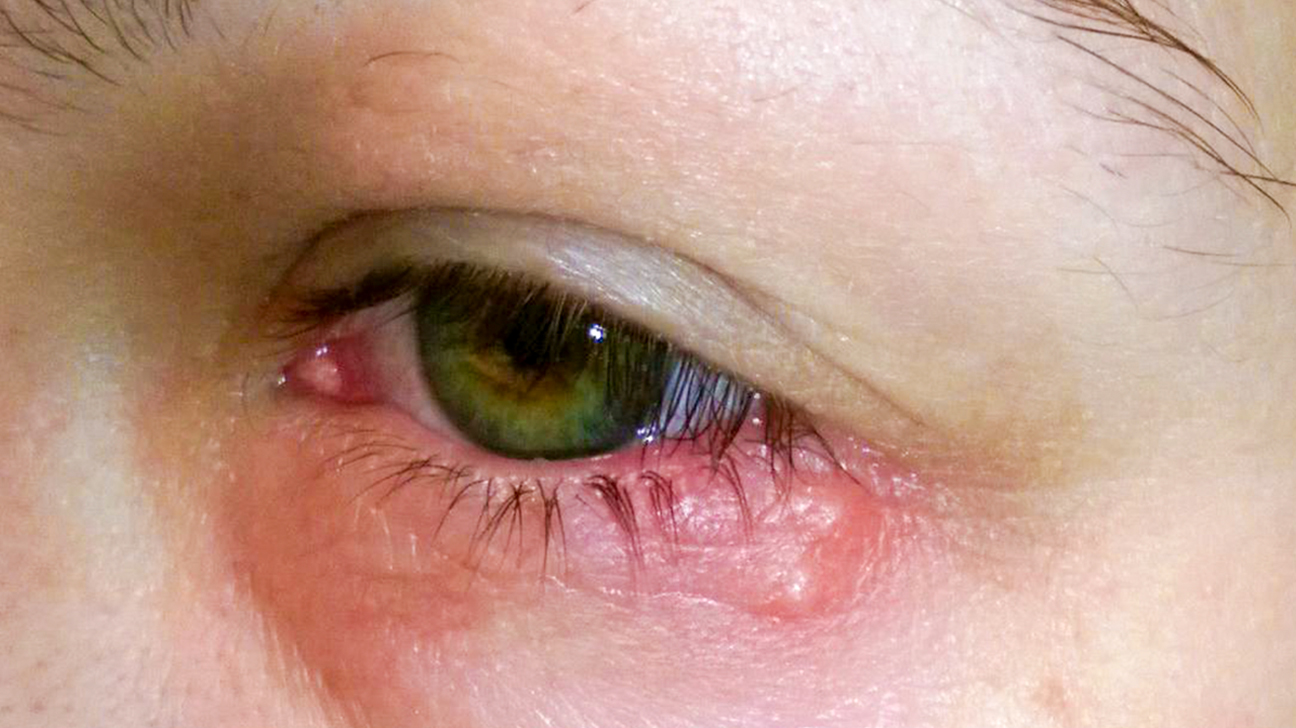
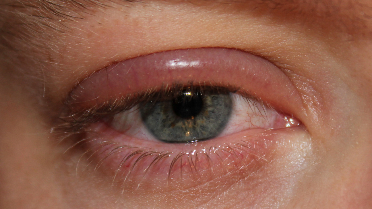
List Of Eye Diseases With Pictures
The eye is a very delicate structure, Eye Anatomy is very complex if there is any problem occur in any part of the eye following diseases can occur:
Picture of Glaucoma

Picture of Cataracts

Age-Related Macular Degeneration

Retinal Detachment

Bacterial Conjunctivitis (Pink Eye)

Uveitis

Eye Allergies

Sty (Stye)

Keratoconus

Chalazion (Eyelid Cyst)

Corneal Ulcer

Strabismus (Crossed Eyes)

How The Eye Works Worksheet
According to the Eye Anatomy following worksheet is designed:
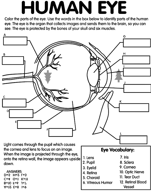
How Does The Eye Work Step By Step?
As we know Eye Anatomy is complex, but it’s functioning is very easy to understand as described in the picture below:

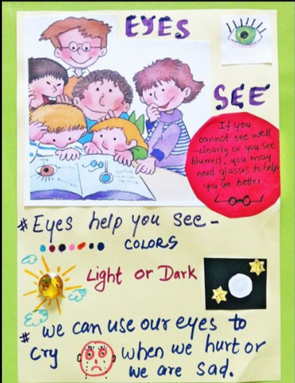
Uses Of Eyes For Kindergarten
Eye Clipart

Eye Cartoon